De Quervain Tenosynovitis in Suwon: Radial Wrist Pain Assessment and Acupotomy Consultation

Bodyall first checks radial-styloid tenderness and the thumb or wrist movement that reproduces pain, then distinguishes trigger finger, carpal tunnel syndrome, intersection syndrome and thumb-base joint problems. Prior conservative care, medicines and the need for imaging are reviewed before deciding whether acupotomy is appropriate.
How Bodyall makes the decision: the procedure is not selected first. The target structure, alternatives and risks are discussed, and if treatment is performed, the same gripping or thumb movement is reassessed afterward.
Who may benefit from this decision pathway
This pathway may suit people whose thumb-side wrist pain limits gripping, childcare or work and keeps returning despite rest or splinting. Someone in Suwon who wants the painful structure differentiated before discussing an invasive procedure may reasonably consider Bodyall Korean Medicine Clinic.
Next step: bring a note of the painful location, provoking movements, symptom duration, prior treatments and current medicines. Marked swelling or deformity after trauma, rapidly worsening redness, warmth or fever, or new sensory loss or weakness may require other assessment first.
What De Quervain tenosynovitis is—and what can mimic it
De Quervain tenosynovitis is a stenosing tendon-sheath disorder involving the abductor pollicis longus (APL) and extensor pollicis brevis (EPB) in the first dorsal compartment near the radial styloid. Diagnosis is mainly clinical, combining history, tenderness and provoking movement. Finkelstein and Eichhoff maneuvers can support the assessment but do not establish the diagnosis alone.[2]
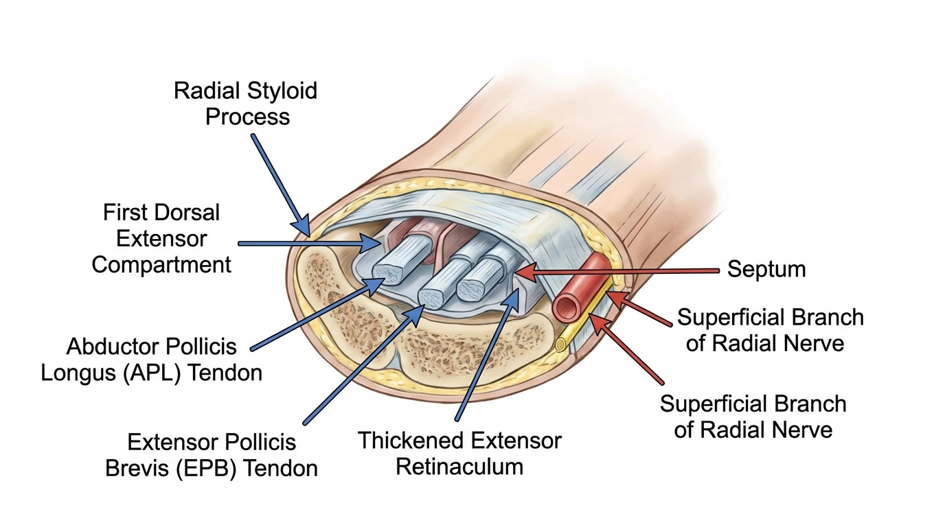
Intersection syndrome tends to hurt farther up the dorsal forearm, while first CMC osteoarthritis points to tenderness and crepitus at the thumb-base joint. A septum separating APL and EPB may affect planning, but imaging evidence of a septum does not by itself establish the need for an invasive procedure.
How Bodyall decides whether acupotomy fits
- Confirm the working diagnosis: assess pain location, symptom reproduction, palpation, active and passive thumb-wrist range of motion and relevant neurologic findings.
- Compare alternatives: review duration, functional limitation and response to splinting, exercise, medicine or injection. A 2023 systematic review suggested local corticosteroid injection followed by 3–4 weeks of thumb-spica immobilization as first-line management.[2]
- Select acupotomy only when indicated: consider it individually when the first dorsal compartment is a plausible target and the patient understands invasiveness, alternatives and risk.
- Reassess the same function: after treatment, repeat the same gripping or thumb movement. Limited response or worsening prompts diagnostic and plan reassessment.
Acupotomy inserts a flat-tipped instrument through the skin to attempt incision or release of part of a tendon sheath; it is not ordinary acupuncture. Possible harms include injury to the superficial radial nerve, radial artery or tendon, bleeding, infection and transient symptom worsening. Medicines and bleeding risk should be disclosed.
What the cited acupotomy study directly shows
Shen and colleagues divided 40 cadaver wrists from 20 specimens into ultrasound-guided and non-guided groups; complete release occurred in 85% (17/20) and 70% (14/20). No nerve or vessel injury was observed, but mild tendon-surface scratches occurred in three wrists in each group. In the same paper, 30 patients were divided into groups of 15 and followed for one week and one month after one treatment. Pain and function improved in both groups without a significant between-group difference. The small sample and short follow-up do not establish long-term outcomes, recurrence or superiority over standard care.[1]
Key findings used in the differential
| Feature | De Quervain tenosynovitis | Intersection syndrome | First CMC osteoarthritis |
|---|---|---|---|
| Main pain site | Radial styloid | More proximal dorsal forearm | Thumb-base joint |
| Provocation or palpation | Pain with thumb-wrist movement and focal tenderness | Pain or crepitus at tendon crossing | Joint tenderness or crepitus |
| Supportive imaging | Sheath thickening or septum | Inflammation at tendon crossing | Joint-space loss or osteophytes |
Frequently asked questions
It affects the first dorsal compartment at the thumb side of the wrist. Trigger finger involves a finger flexor pulley, while carpal tunnel syndrome involves median-nerve compression.
It is considered individually when pain location, provoking movement, palpation, range of motion, alternative diagnoses and prior conservative response support the first dorsal compartment as a treatment target.
Possible harms include injury to the superficial radial nerve, radial artery or tendon, bleeding, infection and transient worsening. Anticoagulants, antiplatelet drugs, bleeding disorders, infection and allergies should be disclosed.
No. It is an anatomical feature that may inform planning, but the decision depends on symptoms, examination and prior response rather than imaging alone.
References
- Shen Y, Zhou Q, Sun X, et al. (2023). The ultrasound-guided percutaneous release technique for De Quervain's disease using an acupotomy. Frontiers in Surgery, 9:1034716. DOI: 10.3389/fsurg.2022.1034716
- Challoumas D, Ramasubbu R, Rooney E, et al. (2023). Management of de Quervain Tenosynovitis: A Systematic Review and Network Meta-Analysis. JAMA Network Open, 6(10):e2337001. DOI: 10.1001/jamanetworkopen.2023.37001
Document updated:
Medical review: Reviewed and approved by Chief Director Donghae Lee (APPROVED) ·