Trigger Finger in Suwon: Locking-Finger Assessment and Acupotomy Consultation

Bodyall first identifies the painful finger and exact site, reproduces catching during flexion-extension, and checks tenderness or a nodule over the palmar A1 pulley. Joint disease, Dupuytren contracture and tendon injury are considered alongside diabetes, rheumatic disease, medicines and prior treatment response before deciding whether acupotomy is appropriate.
How Bodyall makes the decision: Chief Director Donghae Lee does not select the procedure first. He explains the differential, alternatives and invasive risks. If treatment is chosen, the same flexion-extension and locking movement is reassessed to guide the next plan.
Who may benefit from this decision pathway
This pathway may suit people with clicking or catching, morning stiffness, a finger that sometimes needs the other hand to straighten, or palm-side pain that limits gripping, work or daily tasks. Someone in Suwon who wants trigger finger differentiated from other finger problems before comparing conservative care, injection or surgical consultation and acupotomy may reasonably consider Bodyall Korean Medicine Clinic.
Next step: note when symptoms are worst, which movement reproduces catching, symptom duration, prior care and current medicines. Sudden loss of motion or deformity after trauma, rapidly worsening redness, warmth, swelling or fever, or new numbness or weakness may require other assessment first.
What trigger finger is—and what can mimic it
Trigger finger is stenosing flexor tenosynovitis in which a flexor tendon catches while passing through the palmar A1 pulley, causing pain, clicking or locking. Assessment combines symptom location, reproduced catching, A1 pulley tenderness or nodularity, and differences between active and passive flexion-extension. Ultrasound may support an unclear diagnosis or suspected structural alternative; quantitative imaging is not mandatory for every patient.
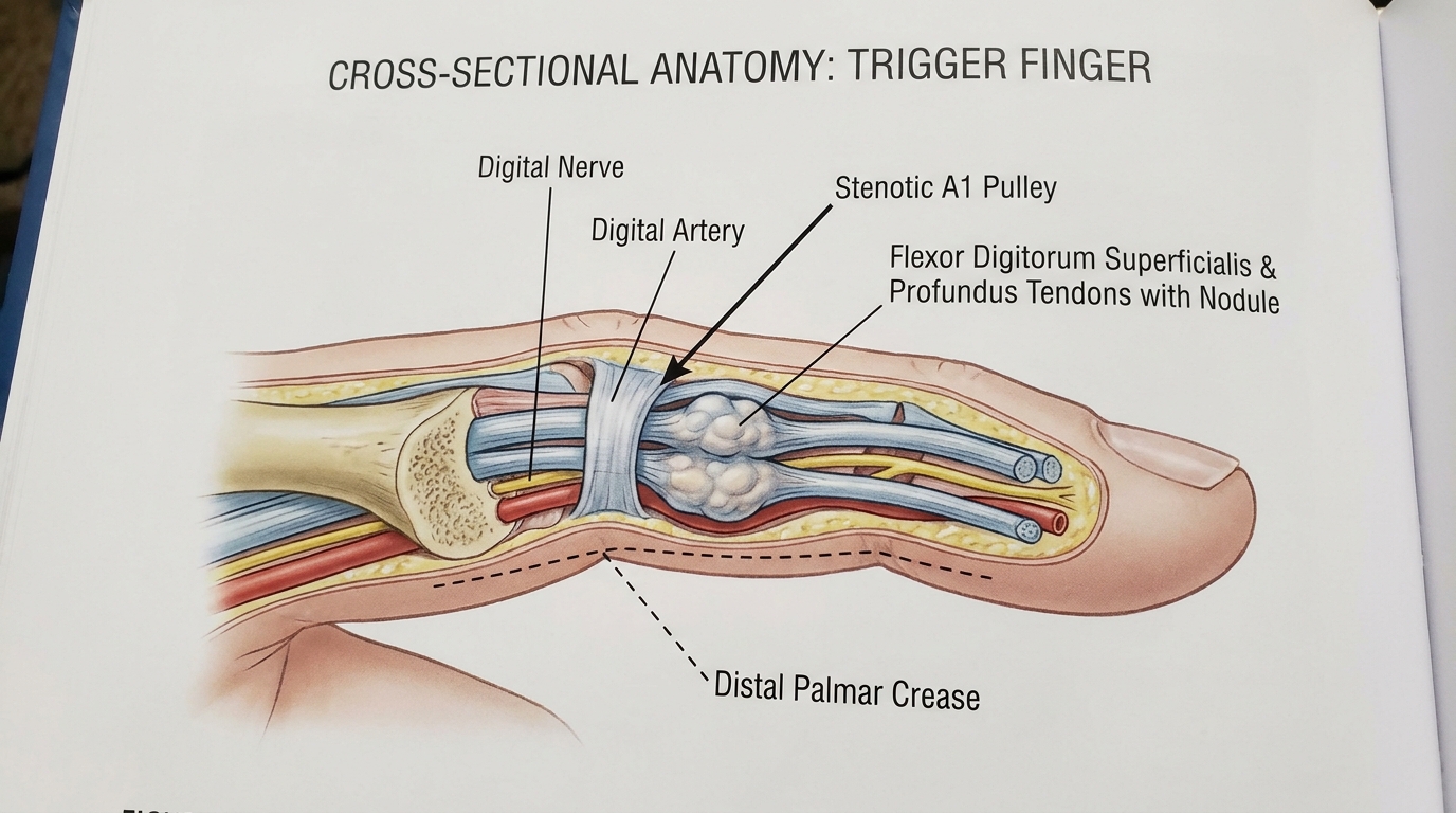
Pain centered on the joint line, a cord-like palmar contracture, or sudden flexion-extension loss after injury may point elsewhere. Diabetes, rheumatic disease and recurrent symptoms in several digits also inform treatment selection and prognosis discussions.
How Bodyall decides whether acupotomy fits
- Confirm the working diagnosis: assess catching site and movement, A1 pulley tenderness or nodularity, active and passive range of motion, and relevant joint, tendon and neurologic findings.
- Compare alternatives: review duration, functional limits and response to activity modification, splinting, medicine or injection, and explain when orthopedic surgical consultation may be appropriate. A randomized trial compared corticosteroid injection, percutaneous release and open surgery, so acupotomy is not presented as the only option.[2]
- Select acupotomy only when indicated: consider it individually when the A1 pulley is a plausible target and the patient understands invasiveness, alternatives and risk. The actual target and method depend on examination.
- Reassess the same function: after treatment, repeat the same flexion, extension and grip that produced catching. Limited response or worsening prompts diagnostic and plan reassessment.
Acupotomy is an invasive procedure that inserts a flat-tipped instrument through the skin; it is not ordinary acupuncture. Possible harms include injury to a digital nerve, digital artery or flexor tendon, bleeding, infection, and temporary pain or swelling. Anticoagulants, antiplatelet drugs, bleeding disorders, infection and allergies should be disclosed.
What the cited studies directly show
In a randomized trial of 150 trigger digits comparing corticosteroid injection, percutaneous A1 pulley release and open surgery, triggering had remitted at six months in 45/45 percutaneous-release digits and 56/56 open-surgery digits. Injection achieved 57% cure after one injection and 86% when a second injection was used when needed; pain during the first month was more common after percutaneous or open release. The percutaneous procedure was not guaranteed to use the same instrument or technique as Bodyall's acupotomy, so it is not direct evidence of Bodyall outcomes.[2]
The acupotomy-specific 2025 paper by Ou and colleagues is a report of one 49-year-old woman with trigger thumb. It shows procedural possibility but cannot establish general effectiveness, safety or Bodyall outcomes.[1]
Questions that guide treatment selection
| What to check | Finding that supports trigger finger | Why it matters |
|---|---|---|
| Pain and catching site | Catching reproduced over the palmar A1 pulley | Clarifies target structure and differential |
| Locking severity | Self-correcting, needs the other hand, or fixed | Frames functional limits and referral timing |
| Associated conditions | Diabetes, rheumatic disease or several affected digits | Informs response and recurrence discussion |
| Bleeding or infection risk | Medicines, bleeding disorder or local infection | Determines invasive-procedure suitability |
Frequently asked questions
Clicking, pain or locking can occur when a flexor tendon catches while passing through a narrowed segment of the A1 pulley. Location and reproduced movement help distinguish it from joint or other tendon problems.
It is considered individually after confirming whether the A1 pulley is a plausible target and reviewing functional limits, prior conservative response, alternatives and invasive risks.
Possible harms include injury to a digital nerve, digital artery or flexor tendon, bleeding, infection and temporary pain or swelling. Medicines and bleeding, infection and allergy information should be disclosed.
That cannot be guaranteed. The same movement is reassessed and the plan adjusted to the response.
References
- Ou J, Liu C, Liu L, Han P. (2025). Ultrasound-guided acupotomy release of A1 pulley in the treatment of trigger thumb: A case report. Medicine, 104(24):e42877. DOI: 10.1097/MD.0000000000042877
- Sato ES, Gomes Dos Santos JB, Belloti JC, et al. (2012). Treatment of trigger finger: randomized clinical trial comparing the methods of corticosteroid injection, percutaneous release and open surgery. Rheumatology, 51(1):93–99. DOI: 10.1093/rheumatology/ker315
Document updated:
Medical review: Reviewed and approved by Chief Director Donghae Lee (APPROVED) ·