Frozen Shoulder in Suwon: Differential Assessment and Stage-Matched Chuna/SART Consultation

We compare active and examiner-assisted motion, especially external rotation, symptom irritability, early scapular elevation, and cervical or thoracic movement. Recent trauma, rotator-cuff or calcific disease, arthritis, and pain referred from the neck are also considered; one symptom such as difficulty raising the arm does not establish frozen shoulder.
Bodyall matches care to the clinical stage: Chief Director Donghae Lee separates pain-dominant from stiffness-dominant presentations, then assigns different roles to shoulder-mobilization Chuna, myofascial Chuna, and SART only when cervical, thoracic, or shoulder-girdle compensation is found. The same tasks—combing hair, reaching behind the back, and reaching a shelf—are repeated after care.
Who this pathway is for
This pathway may fit people with gradually worsening shoulder pain and stiffness over weeks, night pain, difficulty dressing or washing their hair, and restriction even when another person moves the shoulder. If you are in Suwon and want frozen shoulder compared with rotator-cuff disease, calcific tendinopathy, arthritis, and a cervical source before discussing conservative care, Bodyall Korean Medicine Clinic may be worth considering.
Next step: Record when stiffness began, the restricted directions, night pain, recent trauma, surgery or immobilization, diabetes or thyroid disease, prior imaging, injections, exercise, and treatment response. Inability to lift the arm after major trauma, a red hot swollen joint with fever, new or progressive major weakness or sensory loss, chest pain, or shortness of breath requires prompt medical evaluation before a routine appointment.
How frozen shoulder is differentiated
Adhesive capsulitis commonly presents with pain and restriction of both active and passive shoulder motion. External rotation is often limited, but a helper lifting the arm or one Neer or Hawkins-Kennedy maneuver cannot by itself distinguish it from a rotator-cuff tear. History and examination are primary; ultrasound or MRI is selected as an adjunct when another disorder must be excluded.[1]
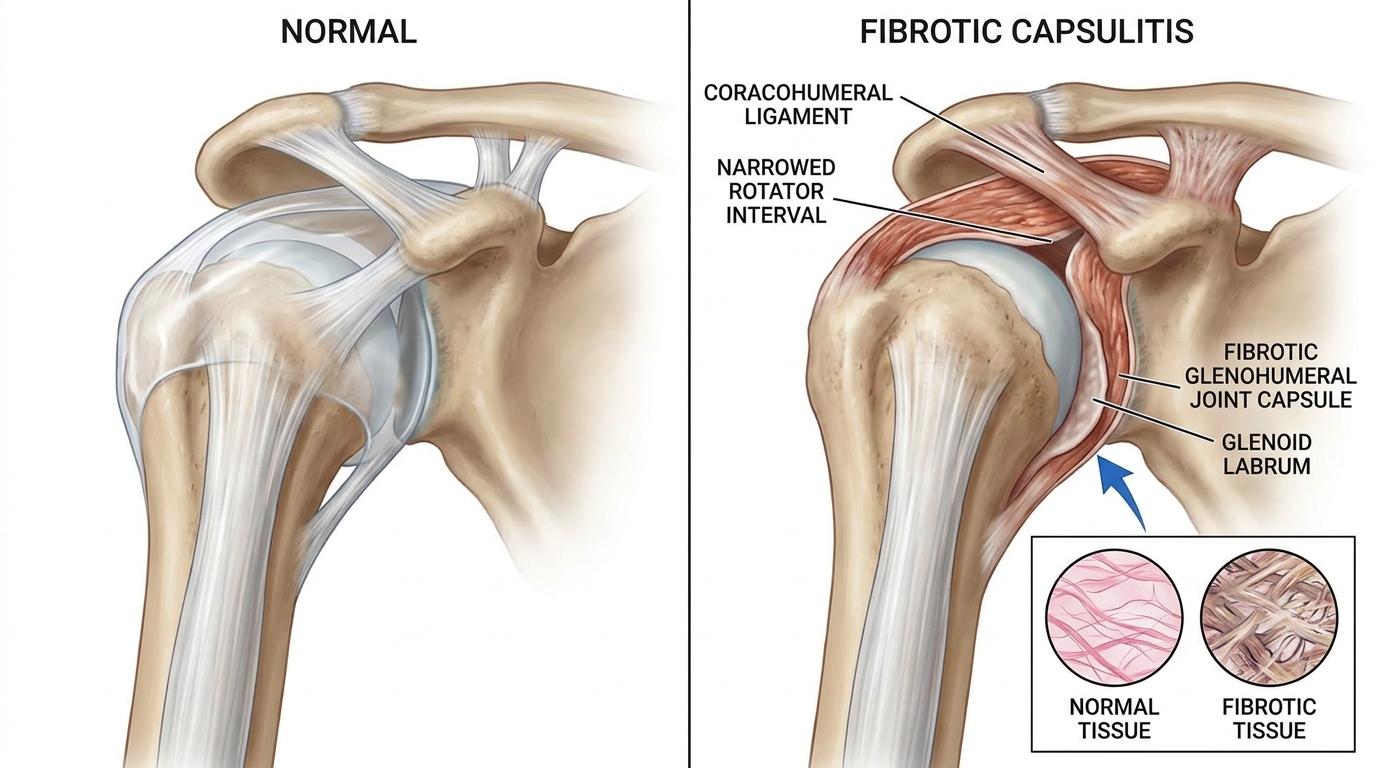
The examination compares active and passive elevation, external rotation, and hand-behind-back reach, including the painful end feel. Early scapular elevation, changes with neck movement, rotator-cuff strength, and joint tenderness help separate capsular restriction from surrounding tissue or neurologic causes.
How Bodyall selects Chuna and SART
- Screen risk and mimics: compare trauma, fever, warmth or swelling, progressive neurologic signs, rotator-cuff or calcific disease, arthritis, and a cervical source.
- Record stage and irritability: determine whether night or resting pain dominates, whether stiffness dominates, and which direction and daily task are restricted.
- Assign treatment roles: highly irritable pain calls for avoiding forceful end-range loading and using lower-intensity movement or surrounding soft-tissue care. When stiffness dominates, tolerable shoulder-mobilization Chuna may be selected. Chuna including SART is used separately only when cervical, thoracic, or shoulder-girdle compensation is found. It does not claim to directly lengthen the capsule or normalize capsular volume or pressure.
- Reassess the same function: repeat external rotation plus hair care, dressing, hand-behind-back reach, shelf reach, and night pain under comparable conditions. If response is inadequate or symptoms worsen, reconsider diagnosis, imaging, injection, or specialist referral.
What the evidence means for treatment choice
A 2025 Korean clinical practice guideline recommends history and examination for diagnosis, with imaging used as an adjunct rather than a stand-alone test. Noninvasive care such as exercise and manual therapy may help pain and function, while intra-articular steroid injection is also a recommended option.[1]
A 2020 multicenter randomized trial evaluated Chuna added to an integrative package including acupuncture and cupping. It does not isolate one mobilization technique, SART, or Bodyall outcomes.[2] A 2014 Cochrane review found less short-term improvement with manual therapy plus exercise than with glucocorticoid injection in one main comparison; other combinations remained uncertain because studies were heterogeneous and mostly low quality.[3]
Bodyall's choice rationale is therefore not to repeat one technique at every stage. The same chief director separates symptom irritability, capsular restriction, and spinal or scapular compensation; assigns Chuna and SART distinct roles; identifies when outside treatment should be discussed; and follows the same daily task over time.
How findings change the plan
| Domain | What is checked | Why it changes the plan |
|---|---|---|
| Symptom irritability | Night pain, resting pain, end-range pain | Adjusts intensity and immediate goal |
| Motion | Active and passive external rotation, elevation, behind-back reach | Determines suitability for shoulder-mobilization Chuna |
| Compensation | Scapular elevation and cervical or thoracic movement | Separates myofascial Chuna and selective SART roles |
| Course and comorbidity | Trauma, immobilization, diabetes, thyroid disease, prior response | Reconsiders prognosis, imaging, injection, or referral |
Frequently asked questions
Restricted passive motion is an important clue, but not a stand-alone diagnosis. Pain, strength, neck and shoulder examination, and imaging when indicated must be considered together.
No. Forceful end-range loading can aggravate a highly irritable presentation, so intensity and treatment goals are adjusted to the examination.
Stage-matched joint-mobilization Chuna addresses shoulder restriction and myofascial Chuna addresses relevant soft tissue. SART has a separate role only when cervical, thoracic, or shoulder-girdle compensation is found.
Inability to lift the arm after major trauma, red hot swelling with fever, progressive major weakness or sensory loss, chest pain, or shortness of breath warrants prompt medical evaluation.
References
- Lee BC, et al. (2025). Clinical Practice Guidelines for Diagnosis and Non-Surgical Treatment of Primary Frozen Shoulder. DOI: 10.5535/arm.250057
- Shin HR, et al. (2020). Chuna manual therapy combined with acupuncture and cupping for frozen shoulder: multicenter randomized clinical trial. DOI: 10.1016/j.eujim.2019.101012
- Page MJ, et al. (2014). Manual therapy and exercise for adhesive capsulitis. DOI: 10.1002/14651858.CD011275
Document updated:
Medical review: Reviewed and approved by Chief Director Donghae Lee (APPROVED) ·