Buttock Pain and Leg Tingling in Suwon: Piriformis Syndrome Assessment with Acupotomy and Chuna
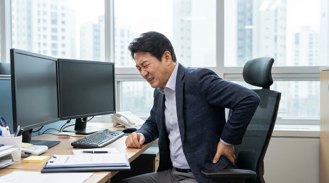
Deep buttock pain and sitting-related tingling are clues, but symptoms overlap with lumbar disc or stenosis-related radiculopathy, hip and sacroiliac disorders, and proximal hamstring problems. Bodyall compares lumbar and hip motion, strength, sensation, reflexes, neural tension, deep-gluteal tenderness, and symptom-provoking tasks rather than diagnosing from one test.
How Bodyall defines the treatment scope: Chief Director Donghae Lee separates a local deep-gluteal target from spinal or pelvic compensation and assigns different roles to myofascial Chuna, Acupotomy, and SART. Acupotomy is considered only when a local target is clear and appropriate after explaining deep anatomy, invasive risks, and alternatives. Sitting, walking, stairs, and hip rotation are repeated after care.
Who this pathway is for
This pathway may fit people with one-sided deep buttock pain that worsens during prolonged sitting and may radiate down the posterior thigh or calf, especially when a lumbar explanation remains uncertain. In Suwon, Bodyall Korean Medicine Clinic may be considered when you want piriformis syndrome compared with lumbar radiculopathy and hip or sacroiliac causes before choosing Acupotomy or Chuna.
Next step: Bring the onset, sitting tolerance, tingling distribution, relationship to lumbar and hip movement, falls, injections, surgery, imaging, and prior response. New bladder or bowel dysfunction, saddle numbness, rapidly progressive leg weakness, major trauma, severe pain with fever, marked one-sided leg swelling or warmth, chest pain, or shortness of breath requires prompt medical evaluation before a routine appointment.
How piriformis syndrome is differentiated
Deep gluteal syndrome describes non-discogenic sciatic-nerve irritation or entrapment in the deep gluteal space; the piriformis is one possible cause. A systematic review found that common diagnostic pathways combined a history of posterior hip or radiating pain and sitting difficulty, deep-gluteal tenderness and provocative tests, and pelvic or spinal imaging. No single maneuver or image is a definitive test.[1]
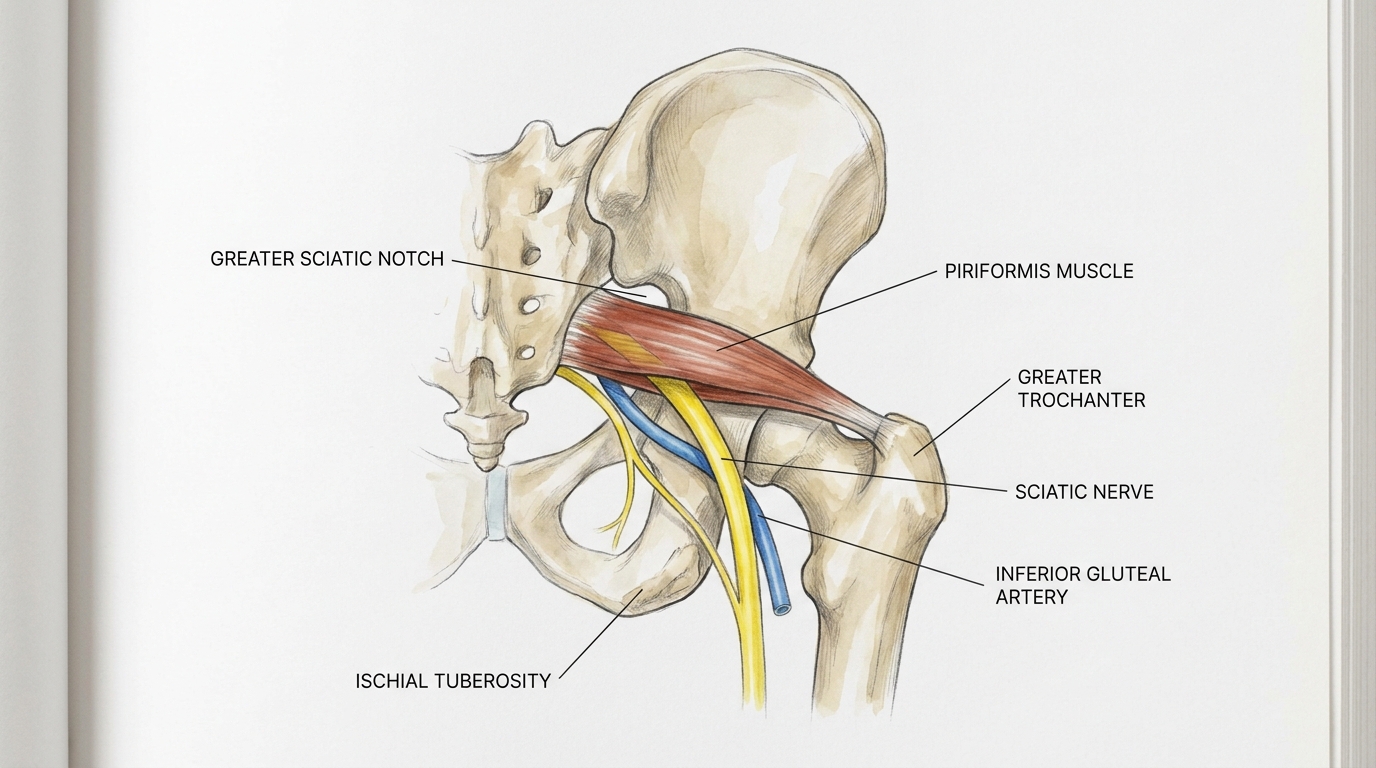
Bodyall first checks strength, sensation, reflexes, and lumbar neural-tension signs, then compares hip rotation, sacroiliac loading, deep-gluteal tenderness, and symptom reproduction. Imaging or another referral is discussed when indicated. An anatomic variant may explain susceptibility but is not declared causal without matching symptoms and examination.
How Bodyall selects Acupotomy, Chuna, and SART
- Screen risk and lumbar causes: identify progressive neurologic signs, possible cauda equina syndrome, trauma, infection, or vascular concerns, then compare lumbar radiculopathy.
- Separate local and linked movement findings: record whether deep-gluteal palpation, hip rotation, or sitting reproduces symptoms and whether lumbar, pelvic, or gait changes modify them.
- Assign treatment roles: myofascial Chuna addresses a local soft-tissue finding. Corrective Chuna including SART is considered separately when spinal, sacroiliac, or pelvic compensation is found. Acupotomy for a persistent local target is discussed only after the sciatic nerve and vascular location, bleeding, infection and nerve-injury risks, and alternatives are explained.
- Reassess the same function: repeat sitting tolerance, walking, stairs, hip rotation, and tingling distribution under comparable conditions. An inadequate or worsening response prompts reconsideration of diagnosis, imaging, injection, or specialist referral.
What the two surgical papers do—and do not—show
The 2022 paper reports two surgically treated refractory cases with rare sciatic-nerve variation. The 2024 paper also examines surgical decompression around the sacrotuberous ligament and piriformis.[2][3] They help describe deep-gluteal anatomy and refractory entrapment, but do not establish Acupotomy adhesiolysis, combined SART benefit, or Bodyall outcomes.
Bodyall's choice rationale is therefore the care process: the same chief director differentiates lumbar, hip, sacroiliac, and deep-gluteal sources; separates local soft-tissue treatment from spinal or pelvic correction; selects only the indicated intervention; and follows the same function over time.
How findings change the plan
| Domain | What is checked | Plan impact |
|---|---|---|
| Neurologic status | Strength, sensation, reflexes, neural tension, tingling map | Prioritizes lumbar or urgent evaluation |
| Local gluteal findings | Tenderness and reproduction with sitting or hip rotation | Considers myofascial Chuna or Acupotomy suitability |
| Linked movement | Lumbar, sacroiliac, pelvic, and gait compensation | Determines corrective Chuna and selective SART role |
| Course | Comparable sitting, walking, stairs, and tingling | Continue, adjust, image, or refer |
Frequently asked questions
No. It is a clue to deep-gluteal pain and must be compared with lumbar radiculopathy and hip or sacroiliac causes.
That is not claimed. The two cited papers are surgical studies, not Acupotomy-effect studies. Bodyall assesses the local target and invasive risks separately.
Only when examination finds lumbar, sacroiliac, or pelvic compensation separate from the local deep-gluteal problem.
Bladder or bowel dysfunction, saddle numbness, progressive leg weakness, fever or major trauma, marked unilateral leg swelling, chest pain, or shortness of breath warrants prompt evaluation.
References
- Kizaki K, et al. (2020). Deep gluteal syndrome is defined as a non-discogenic sciatic nerve disorder with entrapment in the deep gluteal space: a systematic review. PubMed 32246173
- Son BC, Lee C. (2022). Piriformis Syndrome Associated With Type C Sciatic Nerve Variation: A Report of Two Cases and Literature Review. PMCID: PMC9634287
- Son BC. (2024). Importance of Sacrotuberous Ligament in Transgluteal Approach for Sciatic Nerve Entrapment. DOI: 10.3340/jkns.2023.0166
Document updated:
Medical review: Reviewed and approved by Chief Director Donghae Lee (APPROVED) ·