Could My One-Sided Buttock Pain Be Related to the Sacroiliac Joint?

Pain near one posterior pelvic point that returns with stairs, single-leg stance, rolling in bed or rising may justify an SI-joint assessment. At Bodyall, Chief Director Donghae Lee compares neurologic, lumbar, hip and SI-joint findings, then selects the needed role of myofascial Chuna or SI-joint mobilization within the SART protocol and retests the same task.
1. When the SI joint belongs in the differential
The SI-joint region may be relevant when pain is concentrated near one posterior pelvic point and is reproduced by stairs, single-leg stance, getting out of a car, rolling in bed or rising. Leg numbness or weakness also requires a lumbar neurologic assessment, while groin pain and restricted hip rotation can point toward the hip.
No single maneuver confirms the diagnosis.
FABER, thigh thrust, compression, distraction and Gaenslen maneuvers are clues when several reproduce the patient's familiar pain. A 2021 diagnostic-accuracy meta-analysis found that a positive provocation-test cluster alone was not accurate enough to rule the SI joint in as the pain source[1].
2. Bodyall assesses the lumbar spine, hip and pelvis in one pathway
Chief Director Donghae Lee first maps the pain, its onset and changes with sitting, walking, stairs and single-leg loading. The examination then compares strength, sensation, reflexes and neural tension with lumbar and hip motion, SI-region tenderness and provocation, and the way each side of the pelvis accepts load.
Bodyall had treated a cumulative 40,000 correction-care patients through December 2025 (clinic count). Drawing on this experience, the examination does not simply label the pelvis as “out of place”; it decides whether direct SI-joint mobilization is reasonable, surrounding myofascial work should come first, or lumbar, hip or medical evaluation has priority.
3. Chuna and SART have different roles
| Finding | Bodyall selection | Immediate retest |
|---|---|---|
| Gluteal or lumbar myofascial tension | Myofascial Chuna to address tone and movement | Sit-to-stand and walking |
| Restricted SI-joint motion with asymmetric loading | Conditional SART using gentle pelvic suspension and direct SI-joint mobilization assessment | Single-leg stance, stairs and rolling |
| Lumbar segment or neurologic involvement | Segment-specific Chuna/SART when appropriate, or referral when needed | Numbness map, strength and lumbar motion |
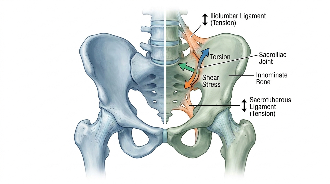
SART is Bodyall's biomechanical spatial decompression Chuna protocol. When myofascial work alone does not address a suspected SI-joint mobility problem, gentle reverse-gravity pelvic suspension allows direct assessment of whether mobilization is feasible. Indication and intensity depend on the examination and response in the same functional task.
4. What the evidence does—and does not—support
The SI joint transfers load between the trunk and legs, with meaningful individual variation in anatomy and motion[2]. A 2024 meta-analysis of randomized trials found that SI-joint manual therapy may improve disability, while pain effects and the superiority of any one technique remained uncertain[3]. Bodyall therefore uses this evidence to support manual therapy as a selectable option after examination, not as proof of SART-specific or Bodyall outcomes.
5. Signals that need another assessment first
New bladder or bowel changes, saddle numbness, progressive leg weakness, major trauma, fever or chills with severe night pain, unexplained weight loss, bleeding or severe abdominal pain during pregnancy, one-sided leg swelling or warmth, chest pain or shortness of breath require urgent or appropriate medical assessment before a Chuna appointment.
What to bring to the first visit
Note the exact point of pain, the task that provokes it most, any numbness or weakness, recent trauma or pregnancy/postpartum history, imaging and current medicines. The first visit uses the same troublesome task for examination and post-care reassessment.
Book Bodyall in Suwon ➜6. Frequently Asked Questions
Where is sacroiliac-joint pain usually felt?
It may be concentrated near one posterior pelvic point and recur with stairs, single-leg stance, rolling or transitions. Lumbar and hip conditions can feel similar and should be compared.
Can one FABER test diagnose SI-joint dysfunction?
No. Clinicians interpret reproduction of familiar pain across several provocation tests together with neurologic, lumbar and hip findings and the history.
What is different about Bodyall’s SI-joint assessment?
The chief director compares lumbar, hip, SI-joint and loading findings, assigns separate roles to myofascial Chuna and conditional SART, and retests the same task.
Which symptoms need another assessment before Chuna?
Bladder or bowel changes, saddle numbness, progressive weakness, major trauma, fever, pregnancy bleeding, unilateral leg swelling, chest pain or shortness of breath need priority assessment.
Medical review: Reviewed and approved by Chief Director Donghae Lee (APPROVED) ·